Overview
The word fracture as used in orthopedic surgery, essentially means “broken bone”. The word fracture does not imply the severity or lack of severity of the break. The most common cause of fractures in small animals involves some form of trauma such as being hit by an automobile. Virtually all bones are susceptible to fracture, but fractures of the long, weight bearing bones (humerus, radius, femur and tibia) and pelvis are most common. Simple non-displaced fractures can be repaired by external coaptation (splints and casts). Complicated and displaced fractures usually require some form of internal fixation such as plates, rods, nails, pins, wires, and screws for stabilization.
The most important aspect of fracture management is to be certain no life threatening problems exist. Fractures, in and of themselves, are almost never life threatening but concurrent thoracic and abdominal trauma may be. Animals that have sustained major trauma should be taken to a general veterinarian or an emergency clinic immediately following trauma. Once the patient is stable and internal injuries have been ruled out or addressed, fracture repair options should be considered.
Fracture classification
Fractures are classified by what bone is involved (example: femur), location of fracture (example: mid-shaft), morphology or configuration (example: spiral), multi-fragments (example: comminuted), if the skin has been penetrated (example: open) and joint involvement (intra-articular). An additional classification system is present for juvenile fractures involving the growth plates (physes). Moreover, complex classification systems exist for specific joint involvement and for open fractures. Fracture classification is useful in communication, fracture planning and prognosis.
Treatment options for fracture repair
Diagnosis of fractures
The diagnoses of fractures are based on physical examination followed by radiographs (X-rays). In some instances, a CT scan is indicated to identify all fracture fragments. CT scans are especially helpful in complicated pelvic fractures and fractures involving joints.
Of course, the first question regarding fracture repair is does the patient require surgery or can the case be well managed with splints, casts and confinement. Non-displaced fractures, especially in young animals are often amendable to external support, also known as external coaptation (splints and casts). External coaptation has pros and cons like any treatment option. The pros are the patient avoids surgery and the initial cost is much less. The cons are splint management, fracture displacement and non-unions. Splints and cast are virtually always problematic in one way or another. Slippage, cast sores, vascular compromise and soiling are the most common problems. Splints and casts must be kept clean and dry and should be changed weekly or every-other week. Animals wearing splints or casts require confinement and Elizabethan collars should be used for the duration of healing (2-3 months on average). That said, there are situation where external coaptation is the best option, but it should not be looked at as an “easy fix”.
Displaced fractures and those involving joints usually require internal fixation. Long bone fractures involving the weight bearing bones of the front or hind limbs, are typically repaired using bone plates, intramedullary nails, pins and wires and/or external fixators.
Plate and Screw Fixation
Plate and screw fixation are arguably the most common form of fracture repair utilized in small animal orthopedics. Plates used for stabilizing fractures are made of surgical stainless steel or titanium and are fixated internally and held in place with bone screws. Newer plate technology involves locking screws that thread into both the bone and plates resulting in greater stability and shorter healing times. Plates come in a plethora of sizes both in thickness and length (figure 1). Figures 2-4 are examples of plate fixation. It is not uncommon to utilize individual bone screws in conjunction with plates.
Figure 1
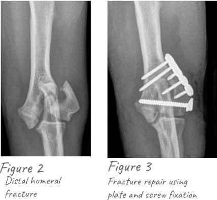
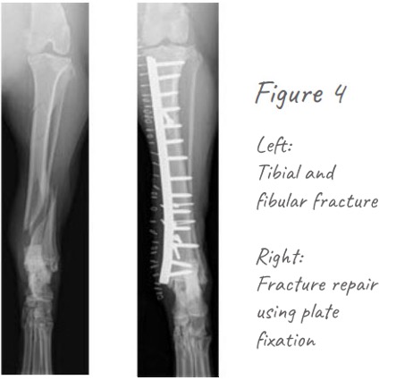
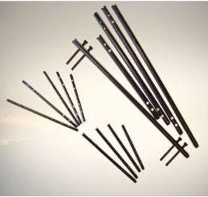
Figure 5: Intrameduary nails
Interlocking, intramedullary (IM) nail fixation
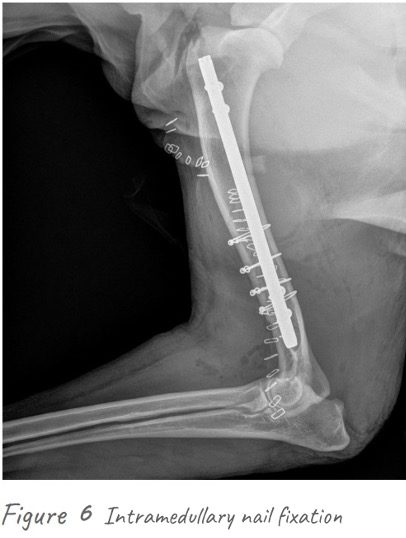
Interlocking, intramedullary (IM) nailing is another established method used to align and stabilize canine fractures (figure 5). Unlike old style IM pins, threaded bolts are placed through holes in the ends of nails to provide rotational stability. This level of strength and stability are not possible with IM pins alone. Nails are inserted into the central canal of long bones of the extremities (e.g. femur, humerus, tibia) (figure 6).
This system offers superior stability for highly comminuted (many pieces) long bone fractures as well as enabling the practice of biological repair (preserving fracture hematoma).
External skeletal fixation
External skeletal fixation has been available to veterinarians for many years (figure 7). There has been a recent resurgence in the use of these devices, as surgeons are finding that minimally invasive reduction of bone fragments combined with rigid fixation provides a more biologic method of fixation for many fractures. External skeletal fixation is also used in conjunction with other repair methods such as IM pinning.
External skeletal fixation involves pins placed percutaneously (through the skin), proximal and distal (above and below) to the fracture fragments and connected to an external frame. External skeletal fixation can be used for various types of fractures and is often used for complex procedures such as angular limb deformity and elbow incongruency correction and distraction osteosynthesis (figures 8 and 9).
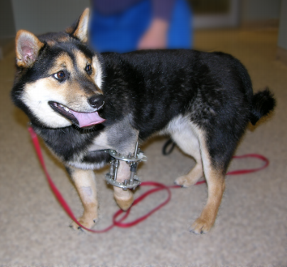
Figure 7
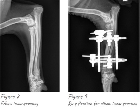
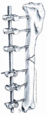
Figure 7 IMEX SK External Skeletal Fixation System Diagram
Recovery and prognosis following fracture repair
Recovery, prognosis and time frame of healing following fracture repair is quite variable depending on the type of fracture and age of the patient. On average, fracture healing progresses gradually over a two to four-month period. During this postoperative period patient activity should be limited. Again, the degree of restrictions depends upon the fracture and fracture repair.
Joint dislocations
Joint dislocations, also called luxations, are common in veterinary orthopedics. Trauma is almost always the culprit although congenital patella luxations and congenital shoulder luxations in toy breed dogs are the exceptions.
In the front limb elbow, luxations are most common but shoulder and carpal (wrist) joints also occur. In the hind limb, hip luxations are by far the most common but again, stifle (knee) and tarsal (ankle) joint luxations also occur. The diagnosis is based on physical examination and radiographs (X-rays). Treatment options are quite variable depending on the joint involved. Closed, non-surgical reduction is possible for some luxations.
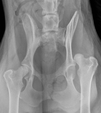
Figure 7
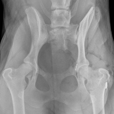
Figure 11: Hip Luxation repaired using a toggle pin technique
Hip or coxofemoral, luxations represent the most common canine joint luxation (Figure 10). Some are amendable to closed, non-surgical reduction, however recurrence is common. Surgical treatment is indicated in dogs with abnormal hip confirmation (hip dysplasia), in situations where a small bone fragments breaks off at the time of luxation and if closed, non-surgical treatment has failed either immediately or weeks later. In dogs with normal hip confirmation, open reductions and stabilization using an “artificial” ligament and toggle pin technique is indicated and carries a very good prognosis. In large breed dogs with abnormal hip confirmation, total hip replacement is the ideal treatment option with an excellent prognosis (Figure 11). In small breed dogs and cats, good to excellent results can be achieved by removing the femoral head (femoral head ostectomy or FHO).
Prices for fracture fixation and joint dislocations are variable based on type of injury and procedure required for repair
Costs for fracture repair and joint luxations vary depending on the extent of the injury and the surgical procedure required. We will provide information about costs for your dog’s case at your consultation. Please feel free to contact us with questions.
At Stitches Veterinary Surgery, we are committed to providing only state-of-the-art, non-compromised pet healthcare. We realize some pet owners may find this level of care relatively costly. However, despite the inherently expensive nature of our work, we are dedicated to providing the highest level of care at the most affordable price possible. We believe if you compare our fees to other specialty practices you will find this true.
